|
|
Fracture Blisters
General Considerations
- Fluid or blood-filled subepidermal vesicles which form over a fracture or severe twisting injury
- Most often in the tibia, ankle and elbow
- May be single or multiple
- May attain sizes in excess of 10cm
- Those at greater risk for formation of the blisters are high impact injuries, those with peripheral vascular disease, diabetes, lymphatic obstruction or a smoking history
Clinical Findings
- May appear as early as 6 hours after injury and usually <48 hours after injury
- They are sterile if intact; once ruptured they can be infected with skin flora
Imaging Findings
- Soft-tissue density, fluid filled masses adjacent to the site of injury
Treatment
- Early immobilization and surgical intervention to repair the fracture (< 24 hrs after injury) reduces the likelihood of their occurrence
- Elevation of injured part
- Use of compression is debatable
Complications
- If the blisters develop prior to operative reduction, they are frequently associated with wound complications
- When the blisters develop following surgery, wound complications are infrequent
- They may lead to a delay in surgery once they form and their presence may alter the operative plan
Prognosis
- Re- epithelialization can take up to 3 weeks

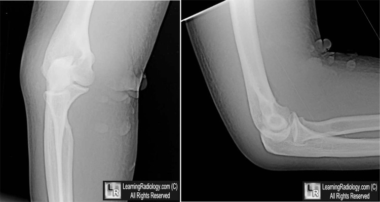
Radiographs of elbow, 10 days after images below

Fracture Blisters. Upper images of the elbow show multiple fluid-filled, soft tissue density vesicles (white arrows)
which occurred shortly after the elbow dislocation three days earlier (lower set of images)
For these same photos without the arrows, click here and here
For more information, click on the link if you see this icon 
Fracture blisters: clinical and pathological aspects. Varela CD, Vaughan TK, Carr JB, Slemmons BK. J Orthop Trauma. 1993;7(5):417-27.
Wheeless' Textbook of Othopaedics
Fracture blisters: a review of the literature. McCann S, Gruen G. Dermatology Nursing.. Dec, 1997
|
|
|
